Breast Carcinoma Types: Ductal, Medullary & Papillary
Breast carcinoma remains one of the most prevalent forms of cancer worldwide, with studies indicating that approximately 1 in 8 women will develop it during their lifetime. Understanding the various breast cancer types—particularly ductal, medullary, and papillary carcinomas—can significantly improve early detection, treatment outcomes, and survival rates.
At the forefront of advanced cancer care, Dr Mathangi J, a Senior Radiation Oncologist with over two decades of expertise, delivers precision-driven treatment strategies tailored to each patient’s diagnosis. With more than 12,000 successful cases and leadership at Gleneagles Cancer Institute, her approach integrates advanced imaging, targeted radiotherapy, and personalized care.
What are breast carcinoma types and why do they matter?
Breast carcinoma types refer to different forms of cancer that originate in breast tissue, each with unique biological behavior, prognosis, and treatment approaches. Identifying the exact type through accurate breast tumor diagnosis allows clinicians to design highly effective, individualized treatment plans.
- Different growth patterns and aggressiveness
- Distinct imaging characteristics
- Varied response to radiation therapy
- Different long-term survival outcomes
Missing the distinction between these types can delay optimal care—something no patient can afford.
What is ductal carcinoma and how is it diagnosed?
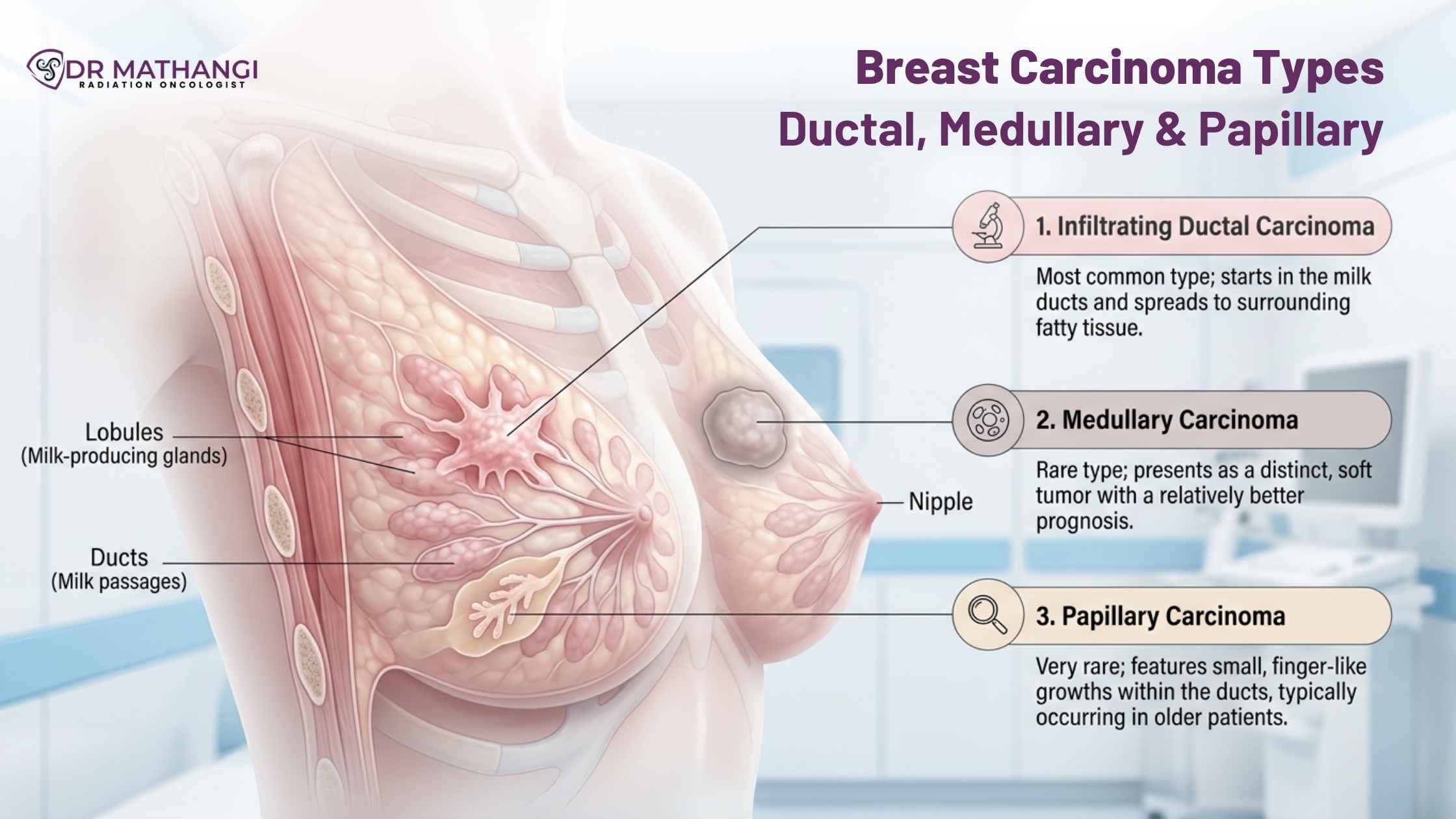
Ductal carcinoma is the most common type of breast cancer, originating in the milk ducts. It can be either non-invasive (DCIS) or invasive (IDC).
Key features of ductal carcinoma
- Accounts for nearly 70–80% of breast cancer cases
- Often detected early through screening
- Can spread beyond ducts if invasive
Role of ductal carcinoma imaging
ductal carcinoma imaging plays a critical role in early detection and staging. Techniques include:
- Mammography
- Ultrasound
- MRI for high-risk patients
These imaging modalities help identify suspicious lesions and guide biopsy decisions.
Mammogram findings carcinoma indicators
Common mammogram findings carcinoma may include:
- Microcalcifications
- Irregular masses
- Architectural distortion
Early identification through these findings can dramatically improve outcomes when treated promptly under expert care.
What is medullary carcinoma of the breast?
Medullary carcinoma is a rare subtype known for its distinct pathological features and relatively better prognosis compared to other invasive cancers.
Understanding medullary carcinoma breast pathology outlines
The term medullary carcinoma breast pathology outlines refers to the structured histological characteristics used by pathologists to identify this cancer type. These include:
- Well-circumscribed tumor borders
- High-grade cells
- Prominent lymphocytic infiltration
Despite its aggressive appearance under the microscope, medullary carcinoma often responds well to treatment, especially when diagnosed early.
Why expert diagnosis matters
Misinterpretation of pathology can lead to overtreatment or undertreatment. With advanced diagnostic precision, Dr Mathangi ensures that patients receive exactly the care they need—no more, no less.
What is papillary carcinoma and how is it different?
Papillary carcinoma is another uncommon type of breast cancer, often seen in older women and typically associated with a favorable prognosis.
Solid papillary carcinoma breast pathology outlines explained
solid papillary carcinoma breast pathology outlines describe the architectural and cellular patterns seen under microscopic examination:
- Well-defined nodules
- Fibrovascular cores
- Low-grade nuclear features
This type is usually slow-growing, but accurate diagnosis is essential to ensure appropriate management.
Clinical behavior and prognosis
Papillary carcinoma tends to:
- Grow slowly
- Have lower metastatic potential
- Respond well to conservative treatment approaches
How is breast tumor diagnosis performed?
Accurate breast tumor diagnosis involves a combination of clinical evaluation, imaging, and pathology.
Step-by-step diagnostic approach
- Clinical examination – Identifying lumps or abnormalities
- Imaging studies – Mammogram, ultrasound, MRI
- Biopsy – Core needle or surgical biopsy
- Pathology analysis – Determining cancer type and grade
This comprehensive approach ensures no detail is overlooked.
Why advanced radiation therapy is critical in breast cancer treatment
Radiation therapy plays a vital role in managing breast cancers, particularly after surgery. Dr Mathangi specializes in cutting-edge techniques such as:
- Stereotactic ablative body radiotherapy (SBRT)
- DIBH gated Radiotherapy
- Image-guided Interstitial Brachytherapy
- RapidArc and IGRT
These technologies allow precise targeting of cancer cells while sparing healthy tissue, significantly reducing side effects.
What cancers require radiation therapy?
Radiation therapy is essential for treating a wide range of cancers, including:
- Breast cancers
- Head and neck cancers
- Lung cancers
- Prostate cancers
- Brain tumors
- Uterine and cervical cancers
Each treatment plan is tailored to the patient’s unique condition, ensuring optimal results.
Why choosing the right oncologist can change everything
Delaying treatment or choosing non-specialized care can lead to disease progression, reduced survival rates, and increased complications. Patients often underestimate the importance of expertise in interpreting imaging, pathology, and treatment planning.
With her extensive training from global institutions and leadership in advanced radiotherapy, Dr Mathangi offers:
- Personalized treatment plans
- Access to cutting-edge technology
- Proven success with thousands of patients
Not choosing the right specialist could mean missing out on life-saving precision care.
How to book an appointment with Dr Mathangi
Taking the first step toward expert cancer care is simple. Patients can submit their contact details via the official form at:
https://drmathangi.com/contact/
Once submitted, her team will promptly schedule and confirm your appointment.
About Dr Mathangi
Dr Mathangi J is a Senior Radiation Oncologist and In-charge at Gleneagles Cancer Institute, Bangalore. With over 20 years of experience and more than 12,000 successfully treated patients, she is a recognized leader in advanced radiotherapy techniques. Her expertise spans breast cancers, lung cancers, brain tumors, and more, making her one of the most trusted names in oncology care in India.
Frequently Asked Questions
What are the main breast cancer types discussed in this article?
The article focuses on key breast cancer types including ductal carcinoma, medullary carcinoma, and papillary carcinoma. Each of these breast cancer types has distinct pathological features, imaging findings, and treatment approaches. Dr. Mathangi provides personalized evaluation and management strategies based on the specific subtype and stage of disease.
How is ductal carcinoma detected through imaging?
Ductal carcinoma imaging plays a crucial role in early detection. Techniques such as mammography, ultrasound, and MRI help identify abnormal tissue patterns. Mammogram findings carcinoma may include microcalcifications or irregular masses. Dr. Mathangi integrates imaging with clinical assessment to ensure accurate diagnosis and timely treatment.
What is medullary carcinoma and how is it identified?
Medullary carcinoma is a rare subtype of breast cancer characterized by well-defined tumor margins and a prominent immune response. References like medullary carcinoma breast pathology outlines help guide diagnosis by highlighting its distinct microscopic features. Dr. Mathangi uses advanced pathology and imaging correlation to confirm the diagnosis and plan treatment.
What should patients know about papillary carcinoma?
Papillary carcinoma is another uncommon form of breast cancer that often presents with unique structural patterns. Resources such as solid papillary carcinoma breast pathology outlines describe its features in detail. Dr. Mathangi ensures careful evaluation through imaging and biopsy to differentiate it from other conditions and provide targeted care.
What are common mammogram findings in carcinoma cases?
Mammogram findings carcinoma may include irregular masses, architectural distortion, or clusters of microcalcifications. These findings often require further evaluation through biopsy. Under Dr. Mathangi’s care, patients receive a comprehensive interpretation of imaging results along with guidance on next steps.
How is a breast tumor diagnosi confirmed?
A breast tumor diagnosi typically involves a combination of imaging, clinical examination, and tissue biopsy. Histopathological analysis confirms the type and grade of cancer. Dr. Mathangi ensures a precise and timely diagnosis, which is essential for selecting the most effective treatment plan.
What treatment options does Dr. Mathangi offer for these carcinoma types?
Dr. Mathangi offers a comprehensive range of treatments including surgery, chemotherapy, radiation therapy, and targeted therapies. The treatment plan is tailored based on the specific breast cancer types, tumor stage, and patient health. A multidisciplinary approach ensures optimal outcomes and patient-centered care.
When should someone consult a specialist like Dr. Mathangi?
It is advisable to consult a specialist if you notice symptoms such as a breast lump, changes in breast shape, or abnormal imaging results. Early consultation allows for prompt diagnosis and management. Dr. Mathangi emphasizes early detection and individualized care for better prognosis.